
Ultrasound means acoustical waves with a frequency above human hearing(20KHz). Ultrasonography is an ultrasound based diagnostic imaging technique. It is mainly used to visualize internal body organs. Sonography is effective for imaging soft body tissues. Structures such as muscles, tendons etc are imaged at a low frequency.

Ultrasound Transducer Working:
In ultrasound transducers mainly two types of conversions takes place
1) Conversion of ac oscillations into acoustic oscillations
2) Conversion of acoustical oscillations back into electrical vibrations
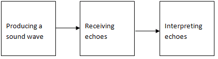
A creation of an image from sound is done in three steps. It is shown in figure below.
Producing a sound wave:
A sound wave is typically produced by a piezoelectric transducer. A piezoelectric element produces a voltage across their two surfaces when deformed. So when the crystal is at rest, no voltage is produced. If the crystal is deformed to the right voltage changes to one polarity and if it is deformed to the left it changes to another polarity.
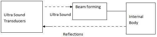
The sound wave is typically produced by a piezoelectric transducer. Strong electrical pulses from the ultrasound machine make the transducer oscillate at desired frequency. The sound is focused to the concerned body part either by the shape of the transducer or by the process called beamforming. This focusing helps the wave travel into the body and focus at a desired depth. The sound wave is partially reflected from the layers between different tissues. Some of the reflections return to the transducer.
Receiving the echoes:
The returning reflections make the transducer to vibrate and these vibrations are reconverted into electrical pulses. The pulses are passed through an ultrasonic scanner and they are processed and transformed into digital images.
Forming the images:
The ultrasonic scanner determines how long it took the echoes to return back to the transducer after the sound wave was sent. It will also determine the strength of the echo. The time taken by the echo to travel back to the probe is measured and it is used to calculate the depth of the tissue interface causing the echo. After this reception of echoes the ultrasonic scanner produces a digital image.