
It is a nuclear medicine imaging technique that produces a 3D image or picture of functional processes in the body. A positron emitting radionuclide (tracer) is introduced in the body. PET system detects a pair of gamma rays emitted from the tracer 3D images of tracer concentration within the body are then constructed by computer analysis. Commonly the biologically active module chosen for Positron Emission Tomography (PET) Scan is fluorodeoxyglucose (FDG) which is an analogue of glucose.
Operation in detail:
Before conducting the scan, a short lived radioactive tracer isotope is injected into the living subject.The tracer is chemically incorporated into a biologically active molecule. The molecule most commonly used for this purpose is FDG. There is a waiting period while active molecule becomes concentrated in tissues of interest. Then the subject is placed in the imaging scanner (usually one hour). During the scan a record of tissue concentration is made as the tracer decays.
As the radioisotope undergoes positron emission decay, it emits a position. Positron is an antiparticle of the electron with opposite charge. The emitted positron travels in the tissue for a short distance. This distance is typically dependent on the isotope. During this travel, the positron loses its kinetic energy until it decelerates to a point. At this point it can interact with an electron. The encounter annihilates both electron and positron producing a pair of annihilation (gamma) photons is detected when they reach a scintillator in the scanning device creating a burst of light. A photomultiplier tube or Si APD will detect this light. The PET technique depends on the simultaneous or coincident detection of the pair of photons moving in opposite direction.
Localization:
The gamma photons are emitted at almost 180 degrees to each other and hence it is possible to localize their source along a straight line of coincidence (called line of response or LOR). In practice the LOR has a finite width. If the resolving time of detectors is less than 500 picoseconds, it is possible to localize the event.
Image reconstruction:
The PET scanner collects a list of coincidence events which represent the simultaneous or near simultaneous detection of annihilation photons by a pair of detectors.
The PET data suffer from scatter and random events much more dramatically than CT. In practice considerable preprocessing of data is required for the correction of random coincidences, estimation and subtraction of scattered photons etc. A common method used to reconstruct images from projections is Filtered Back Projection (FBP). This algorithm is simple and has a low requirement for computing resources. Another method of reconstruction is iterative expectation -maximization algorithms.
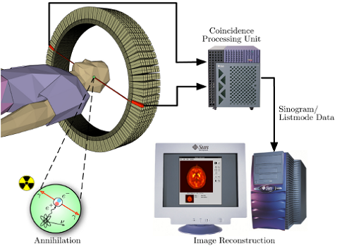
The PET imaging technique is shown briefly in the figure. From the figure we can see that the coincidence detection unit is one of the major units of the PET imaging system.
Attenuation Correction:
Since different LORs must traverse different thicknesses of tissue, the photons are attenuated differentially. As a result the structure deep in the body may be reconstructed falsely. For more faithful representation, the attenuation-corrected images are commonly used.
2D/3D Reconstruction
Modern PET scanners have multiple rings of detectors.
We can use two approaches in reconstructing data from such a scanner.
1) Trace each ring as a separate entity so that only coincidences within a ring are detected. Here the image from each ring can then be reconstructed individually. It is 2D reconstruction.
2) In another method coincidences detected between rings and within rings. The entire volume is reconstructed to form 3D.
Typically Positron Emission Tomography (PET) Scan imaging utilizes a dedicated PET camera system which consists of multiple rings of detectors. The Positron Emission Tomography Scan detectors consist of scintillation crystals coupled with photomultiplier tubes. The scintillation crystals commonly used in PET imaging is BGO (Bismuth Germanium Oxide. The basic concept behind ring design is that the two photons detected in close temporal proximity by two opposed detectors in the ring are likely to have originated from a single annihilation event in the body. Such a simultaneous detection is called coincidence.
Operation in detail:
Before conducting the scan, a short lived radioactive tracer isotope is injected into the living subject.The tracer is chemically incorporated into a biologically active molecule. The molecule most commonly used for this purpose is FDG. There is a waiting period while active molecule becomes concentrated in tissues of interest. Then the subject is placed in the imaging scanner (usually one hour). During the scan a record of tissue concentration is made as the tracer decays.
As the radioisotope undergoes positron emission decay, it emits a position. Positron is an antiparticle of the electron with opposite charge. The emitted positron travels in the tissue for a short distance. This distance is typically dependent on the isotope. During this travel, the positron loses its kinetic energy until it decelerates to a point. At this point it can interact with an electron. The encounter annihilates both electron and positron producing a pair of annihilation (gamma) photons is detected when they reach a scintillator in the scanning device creating a burst of light. A photomultiplier tube or Si APD will detect this light. The PET technique depends on the simultaneous or coincident detection of the pair of photons moving in opposite direction.
Localization:
The gamma photons are emitted at almost 180 degrees to each other and hence it is possible to localize their source along a straight line of coincidence (called line of response or LOR). In practice the LOR has a finite width. If the resolving time of detectors is less than 500 picoseconds, it is possible to localize the event.
Image reconstruction:
The PET scanner collects a list of coincidence events which represent the simultaneous or near simultaneous detection of annihilation photons by a pair of detectors.
The PET data suffer from scatter and random events much more dramatically than CT. In practice considerable preprocessing of data is required for the correction of random coincidences, estimation and subtraction of scattered photons etc. A common method used to reconstruct images from projections is Filtered Back Projection (FBP). This algorithm is simple and has a low requirement for computing resources. Another method of reconstruction is iterative expectation -maximization algorithms.
The PET imaging technique is shown briefly in the figure. From the figure we can see that the coincidence detection unit is one of the major units of the PET imaging system.
Attenuation Correction:
Since different LORs must traverse different thicknesses of tissue, the photons are attenuated differentially. As a result the structure deep in the body may be reconstructed falsely. For more faithful representation, the attenuation-corrected images are commonly used.
2D/3D Reconstruction
Modern PET scanners have multiple rings of detectors.
We can use two approaches in reconstructing data from such a scanner.
1) Trace each ring as a separate entity so that only coincidences within a ring are detected. Here the image from each ring can then be reconstructed individually. It is 2D reconstruction.
2) In another method coincidences detected between rings and within rings. The entire volume is reconstructed to form 3D.
Typically Positron Emission Tomography (PET) Scan imaging utilizes a dedicated PET camera system which consists of multiple rings of detectors. The Positron Emission Tomography Scan detectors consist of scintillation crystals coupled with photomultiplier tubes. The scintillation crystals commonly used in PET imaging is BGO (Bismuth Germanium Oxide. The basic concept behind ring design is that the two photons detected in close temporal proximity by two opposed detectors in the ring are likely to have originated from a single annihilation event in the body. Such a simultaneous detection is called coincidence.